Figure 5

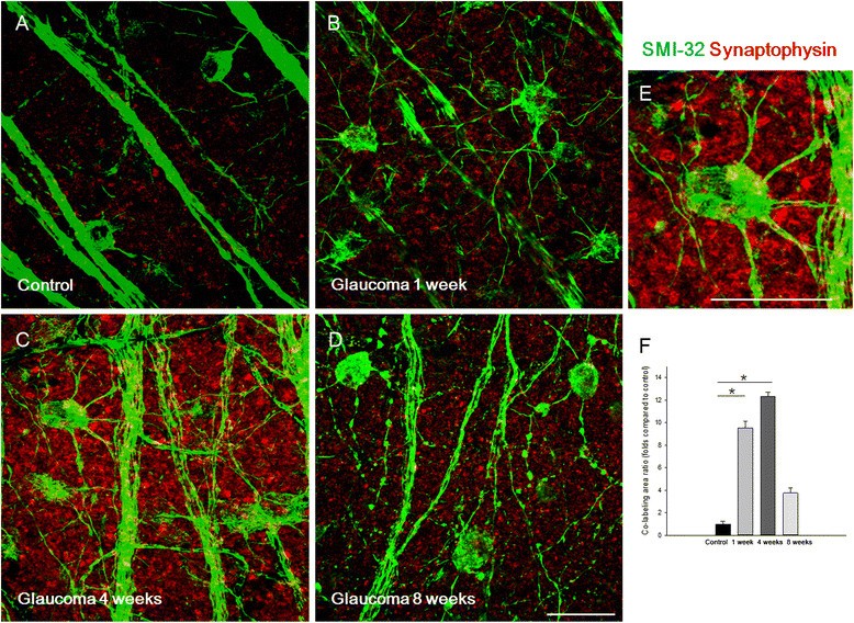
Confocal micrographs of flat-mounted retinas double-stained for synaptophysin and SMI-32, a retinal ganglion cell (RGC) marker, focused on the border of the ganglion cell layer and the inner plexiform layer. SMI-32 staining was seen in both the soma and dendrites of RGCs. Synaptophysin immunoreactivity increased at 1 (B), 4 (C), and 8 weeks (D) after intraocular pressure (IOP) elevation compared with controls (A). SMI-32 immunoreactivity revealed increased and thickened RGC dendrites at 1 (B) and 4 weeks (C), and rounded somas and decreased, beaded dendrites at 8 weeks after IOP elevation (D). Magnification of flat mounts at 4 weeks after IOP elevation (E). Co-staining between synaptophysin and SMI-32 showed that synaptophysin was expressed in the dendrites of RGCs. Co-stained areas significantly increased at 1 and 4 weeks after IOP elevation compared with the control (set to 1.0, F). For flat mount immunohistochemical staining, n=6 for control and n=6 for glaucoma retinas at each time period; total n=24. Scale bars=50 μm.
