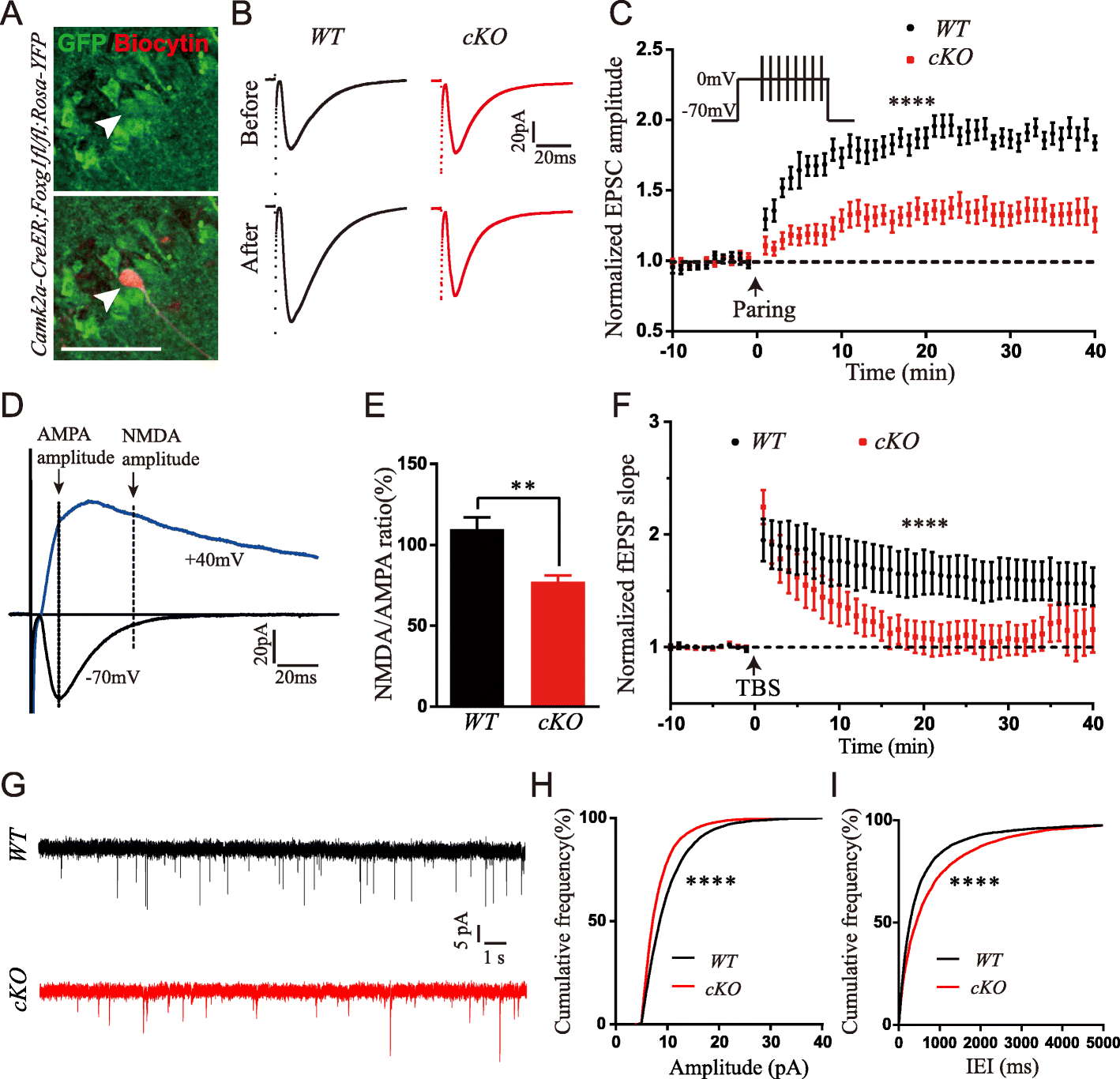
Fig. 3
From: Disruption of Foxg1 impairs neural plasticity leading to social and cognitive behavioral defects

Hippocampal LTP, NMDA receptor activity and synaptic transmission were reduced. a The overlap of post hoc staining of anti-YFP and biocytin verified the precise recording of Foxg1-deficient neurons (Arrow head: upper, YFP+ cells; lower, YFP overlap with biocytin staining. b EPSC amplitude was obviously increased in WT but not cKO mice after paring induction in whole-cell recording (Upper panel, EPSC traces before induction; lower panel, EPSC traces after induction). c LTP was reduced in cKO mice (WT, 15 cells from 6 mice; cKO, 19 cells from 6 mice, F (50, 1632) = 5.021, P < 0.0001; two-way ANOVA, time x group, repeated measure). d Representative traces of AMPA-EPSC (+ 40 mV) and NMDA-EPSC (− 70 mV) from whole-cell recording. e NMDA to AMPA ratio was decreased in cKO mice (WT, 17 cells from 6 mice; cKO, 10 cells from 6 mice, P = 0.0035; t test). f Recordings of field EPSP showed a decreased LTP after TBS induction in cKO mice (WT, 6 slices from 3 mice; cKO, 5 slices from 3 mice, F (43, 387) = 3.657, P < 0.0001, two-way ANOVA, time x group, repeated measure). g Sample traces of spontaneous EPSCs (sEPSC) recorded from hippocampus CA1 pyramidal neurons of WT and cKO. h-i The amplitude was decreased whereas the inter-event intervals were increased in cKO neurons (WT, 33 cells from 6 mice, cKO, 25 cells from 6 mice. Amplitude, P < 0.0001; IEI, P < 0.0001, K-S test). Scale bar, 50 μm. **P < 0.01, ****P < 0.0001
